 |
| Pictured- an echinococcus species. |
Tapeworms are segmented and ribbon like. They have no gut, and nutrients are absorbed across the tegument. This means that the worm relies on the horse for nutrition and renders them obligate parasites. They have an indirect life cycle, which means they have one or more hosts in their development cycle. They are hermaphrodites and are not split into sexes.
Morphology
- The rostellum doesn't always have hooks.
- There are generally 4 suckers, which may also possess hooks.
- An immature segment of the worm is known as a proglottid, and does not contain eggs.
- The genital pore pictured, can be a diagnostic feature in some worms.
- A proglottid which contains eggs is known as the gravid proglottid. This breaks off and sheds into the gut and is released into faeces.
- Tapeworms possess a bi-lateral excretory duct.
- Taenia species have a single lateral excretory (genital) pore.
- Dipylidium Caninum has a double excretory pore in each segment which forms a rice grain shape on both sides.

- The life cycle pictured above shows a typical tapeworm life cycle. The proglottids are shed in the faces of the animal by the adult worm (in this instance a human) and these are the infective stage for the intermediate host. The egg pictured on the right features a hexacanth in the middle which is the embryo or an ochosphere. The outside layer is the thick, brown embryophore.
- The eggs are ingested by the intermediate host which is in this instance the cow. This stage encysts in the host, and is generally referred to as the metacestode stage of the tapeworm.
- The meat of the animal is eaten by the definitive host which is the end stage host and this releases the tapeworm which develops to the adult stage.
Metacestode Terms
 |
| Cysticercus Diagram |
- A cysticercus is most common and the tapeworm is contained in a fluid filled cyst with a protuding protoscolex.
- A coenurus consists of several tapeworms.
- A hydatid cyst consists of more than 100 tapeworms.
- A cysticercoid is a cyst that is solid rather than fluid filled. These generally have an invertebrate intermediate host.
The metacestode has economical significance in a variety of species because of the possible zoonosis to humans if the muscle of the animal is eaten containing these cysts. There are a variety of controls in place in abattoirs to detect these cysts. The site of the cyst is diagnostic to the species of tapeworm. Treatment of these is generally with praziquantel.
Taenia Species
Definitive host generally is the small intestine of carnivores. The adults are non-pathogenic. They have an armed rostellum (hooks) and characteristic eggs.
Taenia Saginata
- Cysticercus in the muscle of beef cows.
- Zoonotic- causes mild pain in humans from hunger and feels unpleasant.
- Control-> meat inspection, cooking or freezing meat and proper sewage disposal.
Taenia Solium
- Eradicated in the UK.
- Highly pathogenic cysticercus stage in pigs and humans.
- Humans ingest eggs-> Poor sanitation or meat inspection. Leads to epilepsy, sub-cutaneous lumps and neural-ocular symptoms.
- Zoonotic.
| Taenia Multiceps- sheep brain. |
Taenia Ovis (muscles of sheep), Taenia Hydatigena (peritoneum and liver fibrosis) and T. Multiceps
- Intermediate host for all= sheep.
- Definitive host= dog.
- Pre-patent period (PPP) which is the time from ingestion to the appearance of eggs in the dog's faeces= 6 weeks.
Taenia Pisiformis (peritoneum) and Taenia Serialis (connective tissue and subcutaneously)
- Intermediate host for all= rabbit.
- Definitive host= dog.
- T.Serialis develops coenurus where the rabbits are kept in runs or on lawns.
Taenia Taeniformis
- Intermediate host= mouse. Liver cysticercus.
- Definitive host= cat. Bell shaped proglottid= diagnostic feature. 6 day ppp. Reinfection is common as it is hard to control access to the intermediate host and metacestode stage.
Echinococcus Species
| E.granulosus in H & E stain |
Echinococcus Granulosus
- Intermediate host= sheep/humans.
- Definitive host= dog (can be as many as 700 tapeworms per dog).
- E.granulosus granulosus= sheep intermediate host.
- E.granulosus equinus= horse intermediate host (not zoonotic).
- Human hydatid disease-> ingestion of eggs. Space occupying cyst in the liver, lungs, bone marrow or brain. Slow growing accompanied by jaundice, cholangitis, abdominal pain, pleurisy and if the cysts rupture- anaphylactic shock. Singular cysts.
- Found in developing countries and also mid wales. Controls are effective only if the control programme is maintained.
Echinococcus Multilocularis
- A big growing concern in the UK.
- Currently found in other countries.
- Highly pathogenic zoonosis.
- Large slow growing cysts that metastasise and spread.
- Difficult to diagnose and treat as impossible to remove cysts.
- Fox-rodent cycle so urban fox population in the UK a concern.
- Human= accidental infection.
Horse Cestodes- Anoplocephalidae family
- Horse= definitive host.
- Orbatid Mite= intermediate host.
- Metacestode stage= cysticercoid.
- Anoplocephala perfolita- found at the Ileo-caecal junction. Possesses short broad segments and lappets behind each of the four suckers.
- Mites live in the soil and hay. They are common and microscopic. These are ingested by the horse. 2 month development to the adult.
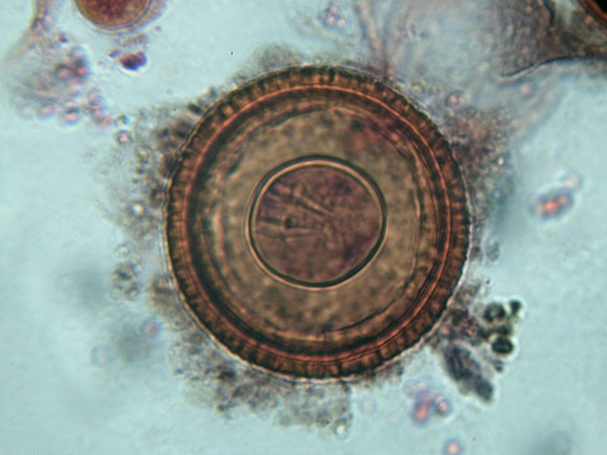
- 'D' shaped egg with pyriform apparatus and onchosphere.
 |
| Egg. |

Pathogenesis
- Spasmodic colic.
- Intussusception.
- Ileal impaction.
- Rupture of the intestine.
Clinical Signs
- Ulceration.
- Unthriftiness.
- Enteritis.
- Colic (spasmodic and ileal impaction).
Epidemiology
- All ages. Below 3 years old more likely to be infected. Peak= autumn/winter. Infection is possible all year round.
Diagnosis
- Faecal egg count= 60% sensitivity (the probability of a positive test).
- ELISA- can be used to detect circulating antigens and have 68% sensitivity and 90% specificity (probability of a negative test). This is the IgGT subtype antibody which is used in response to the 12/13 KDA component of the parasite excretory/secretory antigen. This correlates with infection intensity.
Treatment
- Praziquantel.
- Double douse pyrantel.
Monezia
- Definitive host= sheep, cattle and goat small intestine.
- Intermediate host= cysticercoid in orbatid mite intermediate host.
- Not very pathogenic.
 |
| Dipylidium egg- capsule containing 10+ eggs |
Dipylidium Caninum
- Cat and dog small intestine= definitive host.
- Intermediate host= trichodectes canis (dog lice) or ctenophalides felis (cat flea). A cysticercoid found in the body cavity. They are eaten as the adult dog grooms.
- Proglottids in the dog faeces are motile.
- Cause irritation, but mainly unpathogenic.
- Treatment= praziquantel and ectoparasite control.
No comments:
Post a Comment